Internal marking is a vital step in yielding good surgical results, protecting the patient, and keeping the operating room staff on the same page. Unlike external marking, internal marking uses the application of marks with dedicated surgical markers, dyes, clips, sutures, and other means, inside the body.
Internal surgical marking assists surgeons in ways such as:
- Locating tumors before removing them
- Transplanting organs
- Placing prostheses
- Preserving important blood vessels or nerves
- Avoiding mistakes, which might cause dangerous internal bleeding
- Classifying tissues for histopathological and radiologic tests
This article discusses surgeries where internal tissue marking is essentially a must in achieving a successful outcome.
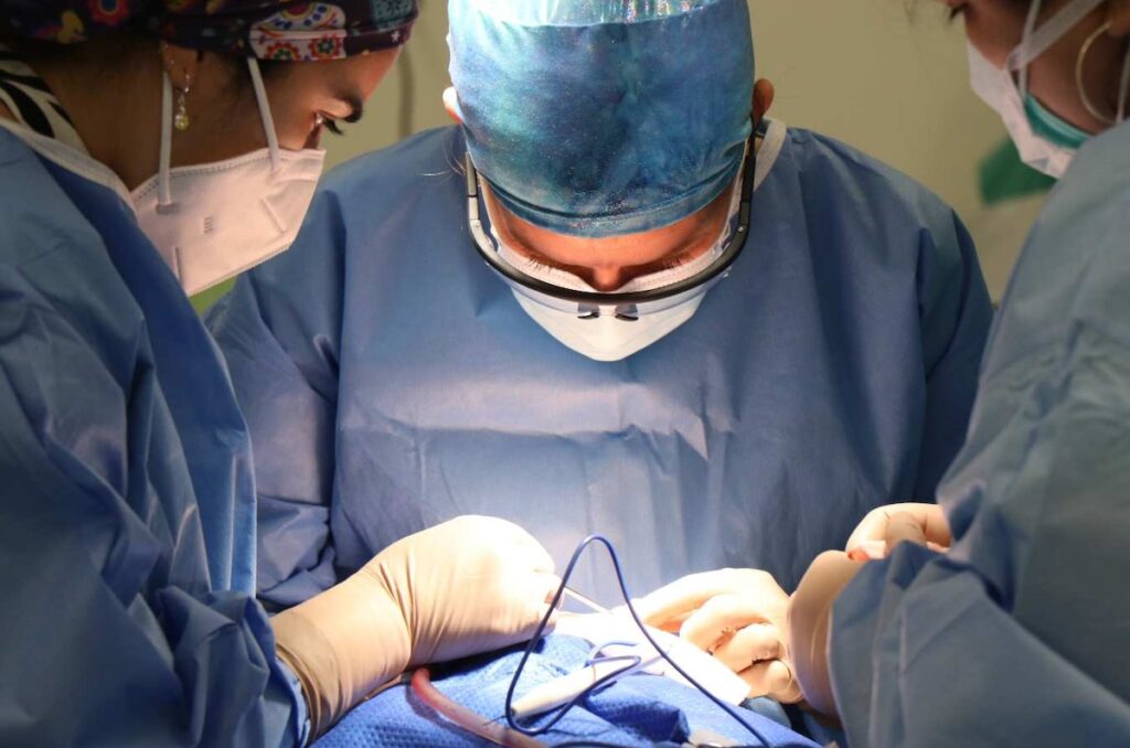
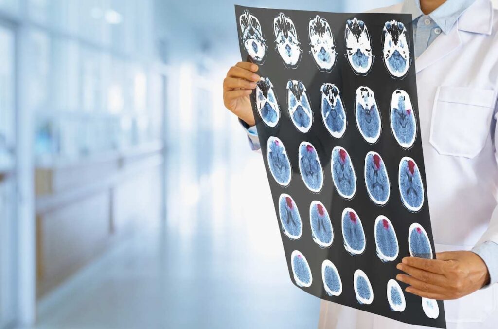
1. Brain tumor removal
Brain tumor removal involves surgically removing tumors from the brain. It’s delicate, as brain tumors often have unclear margins. Brain tumors tend to infiltrate their surroundings, so they need to be demarcated.
Neurosurgeons often choose not to use intraoperative marking to save time. But recent findings show internal marking proves beneficial in:
- Making resections more successful
- Saving healthy tissue
- Identifying critical brain structures
- Navigating within limited spaces
- Improving patient outcomes
One neurological study elaborated on the role of dyes in meningioma surgeries. Meningiomas are the most common primary brain tumors, accounting for 30% of all brain tumors.
According to the study, surgical marking dyes distinguish meningiomas from healthy dural tissue. The studied dyes included:
- 5-aminolevulinic acid (5-ALA)
- Indocyanine green (ICG)
- Fluorescein
Another study evaluated the effect of internal marking on glioma tumor removal. The study suggests that certain dyes (5-ALA or CLR1502) can delineate tissue margins during surgery. These forms of marking can provide a means of removing tumors while preserving healthy tissue.
2. Breast cancer lumpectomy
A lumpectomy is a type of surgery that removes a breast tumor and a margin of surrounding tissue. It’s often preferred to a mastectomy, which is a more radical treatment to fully remove the affected breast.
Breast lumpectomy has a high rate of re-excision. Research shows that about 1 in 4 women must undergo re-excision surgery after a lumpectomy. The reason is partly due to incomplete tumor margins. This is where surgical marking comes into play.
Correctly marking breast cancer margins results in:
- Localizing breast tumors and their extent accurately
- Fewer re-excisions
- More accurate re-excisions
- A lower rate of cancer recurrence
- Conservation of breast tissue
- Improved breast cosmetics
Internal marking during a lumpectomy can be done with:
- Readily available OR instruments, such as foam boards, sutures, and clips
- Intraoperative breast surgery marking pens
- Advanced methods, such as 3D absorbable inks
3. Liver resection (hepatectomy)
Liver resection surgery involves removing a part of the liver containing primary cancer or metastases. It’s another operation where internal tissue marking has proved essential.
Permanent surgical marker pens accurately mark incision sites during liver resection surgeries. These markers can serve as guides for surgeons to access the liver tumor location.
A review from the Annals of Surgery studied the application of indocyanine green (ICG) dye in liver surgery, suggesting it can be useful because it’s a simple and safe dye for laparoscopic and open liver resections, and it can depict hepatic tumors and liver segmentation with precision.
Another study discussed the possible benefits of BL-760, a novel ink for liver surgery. This ink can:
- Effectively delineate the hepatic bile ducts
- Identify bile leaks and minor duct injuries. Bile leaks are, more often than not, missed by surgeons during operations. This mistake can be life-threatening, as it might lead to liver failure and sepsis.
- Show a high TBR (tumor-to-background ratio) fluorescence, which means it can visualize tumors well
- Be removed quickly from the body
- Be reapplied intravenously (unlike ICG)
4. Orthopedic joint replacement (arthroplasty)
A joint replacement surgery aims to restore joint function with prostheses or artificial joints. Prostheses can replace various joints, such as the hip, knee, or shoulder.
To ensure correct prosthesis placement, marking with a surgical marker pen is usually done before and during arthroplasties. Surgeons utilize internal marker pens to mark incision sites and define the extent of bone removal. Gentian violet markers are commonly used, as they’re sterile and have antifungal properties.
But there are some potential dangers with gentian violet that should be kept in mind.
Simple sutures can also be used to mark tissues. For instance, during hip replacement surgery (HTA), surgeons often cut parts of muscle tendons to achieve better access to the hip joint. An example of such a tendon is the piriformis muscle tendon. These cuts provide better access to the joint. Then, they can use sutures to mark the tendons and repair them afterward.
These practices of internal marking can:
- Define the exact amount of bone removal
- Pinpoint the location of the prosthesis
- Ensure alignment and functionality of the prosthesis
- Preserve crucial vessels and nerves (such as the sciatic nerve)
- Prevent damage to muscle
- Prevent future joint problems and re-operations
5. Coronary artery bypass grafting (CABG)
Coronary artery bypass grafting (CABG) is a procedure to restore blood flow to the heart. CABG involves fixing coronary artery blockage using a graft – a substitute vessel from another body part. The most commonly used grafts include the saphenous vein and internal mammary artery
So, how can surgeons ensure they’ve got the graft right where it needs to be during the operation?
Internal surgical marking provides the answer. Internal marking can assist in:
- Finding the correct attachment spot for the graft
- Visualizing the coronary arteries
- Evaluating graft patency
- Identifying anatomical variations
- Dealing with complex CABG surgeries
- Obese or diabetic patients
- Complicated coronary anatomies
- Congenital surgeries
Graft marker pens are widely used before or during the preparation of vein grafts used in bypass surgery. These pens demarcate selected sites and the orientation of the graft. They’re handy, sterile, non-sterile, and typically planned for a single use.
Additionally, various surgical dyes can visualize the coronary arteries. These include the following:
- Indocyanine green (ICG, Spy Agent Green)
- Methylene blue
- Fluorescein
6. Colorectal surgery for cancer
Colorectal surgery is often performed to remove cancers from the colon and rectum. It comprises three main types:
- Polypectomy – Removal of polyps using colonoscopy.
- Local excision – Removal of small cancers from the lining of the colon. This type also uses colonoscopy.
- Colectomy – Partial or complete resection of the colon. A colectomy may be a laparoscopic or an open-type surgery. A colectomy also involves removing nearby lymph nodes.
Studies have shown that internal marking is critical for colorectal surgery. Marking is also a prerequisite for successful outcomes in colorectal surgery.
A case report suggests marking clips can be applied successfully in colorectal cancers. It argues that these clips can:
- Visualize polyps and tumors with a fluorescent laparoscope.
- Help preserve healthy tissues.
- Provide more safety than dyes.
- Be placed only a day before the surgery.
- Avoid the risk of complications associated with “tattoo marking” techniques.
Sutures can also be used intraoperatively to label the main tumor-feeding artery.
“Tattoo marking” is the traditional way of labeling gastrointestinal tumors. This method is widely used in colorectal surgery, too. It utilizes various dyes or inks, like ICG. However, newer studies have shown tattoo marking might increase the risk of punctures and peritoneal injury.
7. Pancreaticoduodenectomy (Whipple procedure)
Pancreaticoduodenectomy is a surgery that removes pancreatic or duodenal tumors. It’s also called the Whipple procedure, for short.
The anatomy around the pancreas and duodenum is complex. Likewise, tumors in the regions are often complex and life-threatening. They might spread close to major organs and blood vessels. Hence, setting accurate resection margins through internal marking is necessary.
Studies show that markers and dyes of various colors, and silk sutures can:
- Outline the tumor’s location
- Help achieve margin-negative resections
A margin-negative resection means no cancer cells lie at the edges of the removed tissue. A margin-negative resection indicates an R0 resection (complete cure).
- Identify vital structures, such as:
- Duodenal parts that are vulnerable to ischemia
- Major vessels, such as the superior mesenteric artery and the portal vein
- Nerve plexuses that line vessels
- Play a crucial role in diagnosing and staging tumors.
Other surgeries where surgical marking is vital
Various novel marking techniques have also been developed for other types of surgeries. Different surgical markers and inks have the potential to contribute in important operations, such as:
- Thyroidectomy
- Renal transplant surgery
- Arteriovenous malformation surgery
