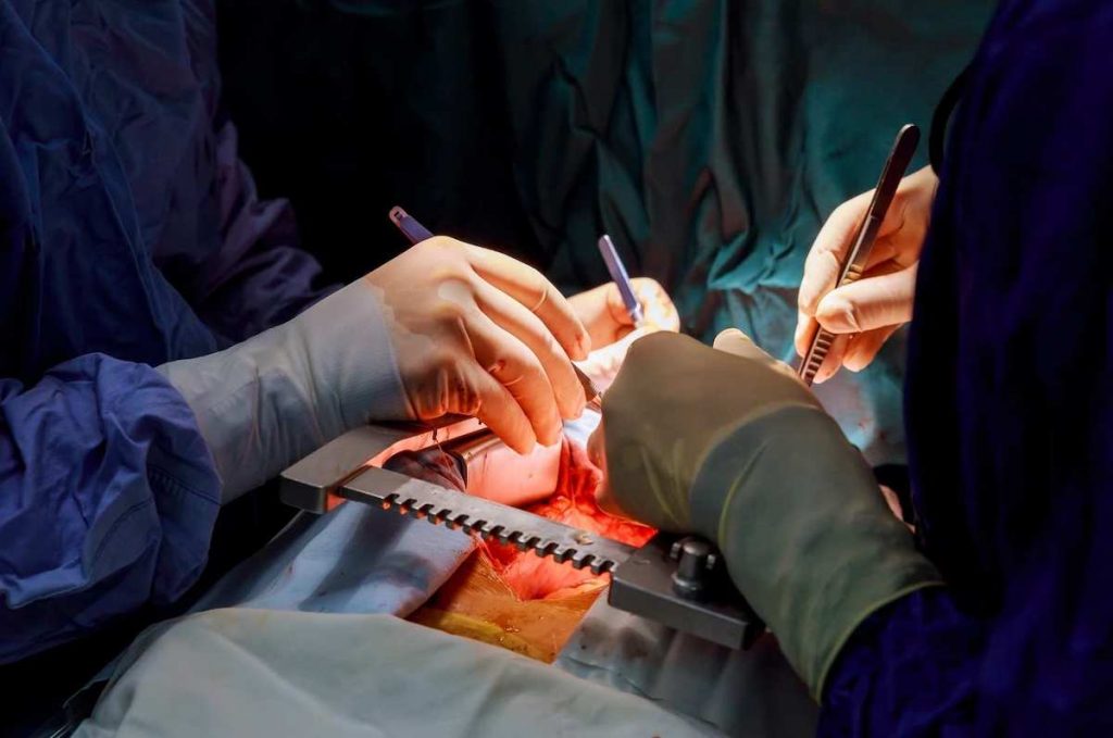
Surgeons employ various types of surgical markers and dyes to mark inside patients during surgery. Internal surgical markings can demarcate tumor margins, help identify and preserve delicate anatomical structures, and facilitate resection and transplantation procedures, among other aims.
Ultimately, they are aimed at more accurate surgery, mitigating risks and potential errors, and safeguarding the patient’s overall well-being. This article reviews some of the main reasons surgeons mark inside patients during surgery.
Tumor margin and biopsy sample identification
Surgical marking is pivotal in oncologic/cancer surgery. Various markers and dyes are frequently employed by oncologic surgeons and pathologists to carry out their respective roles with greater precision, which ultimately benefits the diagnosis and treatment of the patient.
Identifying tumor margins and highlighting specific areas of interest on tissue specimens during surgery is one of the primary applications of internal surgical marking.
Traditionally, gentian violet was used for this purpose, but in contemporary medicine, many vibrant, multicolored surgical dyes have become available. Margin marking is aimed at relating the surfaces of the tumor to their original anatomical locations. This crucial step helps pathologists orient themselves during the histopathologic examination of tumor margins and lets them communicate the precise locations for subsequent surgical resection to surgeons in cases of positive tissue margins.
Overall, this practice significantly diminishes the risk of leaving behind residual cancer cells, thereby greatly minimizing the chances of cancer recurrence.
Surgical dyes are equally valuable in the context of labeling biopsy specimens. Biopsies involve the removal of one or more small tissue samples from an organ of interest for examination and diagnosis. Often, multiple samples are collected from different areas within the same organ or tissue. This is the case with sextant biopsies of the prostate. This procedure involves the removal of tissue samples from six different areas of the prostate.
These core tissue samples are then labeled with blue, black, or green dyes to help histopathologists determine the origins of the sample, especially if they are embedded and analyzed together on the same slide. By dyeing these different “cores,” histopathologic examination can be expedited by allowing the simultaneous examination of multiple samples on the same slide without sacrificing valuable information about the tumor location.
Organ tissue marking
Surgical marking plays a crucial role in organ procurement surgeries, especially when dealing with marking organ laterality. Accurate laterality determination by transplant surgeons ensures structural compatibility between to donor and recipient and prevents the potential complications that may arise from the mislabeling of organs.
The clearest example of the critical role of organ marking occurs during kidney procurement surgery. The identification and harvesting of the correct-sided kidney are of paramount importance during renal transplant. The consequences of incorrectly labeled laterality during this procedure can range from annoyance created by not receiving the expected kidney, to prolonged cold ischemia time, or even the disposal of the received kidney as it cannot be used for the intended recipient.
An example is the United Network for Organ Sharing (UNOS), recorded in 2012 to 2014. Consequently, marking kidney laterality has become mandatory in most renal procurement procedures. This can be accomplished by employing various methods, such as placing a silk tie, prolene suture, or umbilical tape on the distal ureter or distal renal vein, or by placing color-coded vessel loops in the perinephric fat.
Such practices ensure that transplant teams can confidently proceed with procurement procedures while the receiving team can accurately orient and position the organ within the recipient’s body, avoiding costly errors and ensuring successful kidney transplants.
Tissue resection and guidance
Surgical marking can also be used to identify tissues and lesions for resection, greatly improving the precision and success of these interventions. Marking can also be done following tissue removal to mark removal sites for guidance in future interventions.
“Endoscopic tattooing” of colorectal lesions is a good example of using surgical inks and dyes to localize small lesions or previous resection sites. Today, the majority of colorectal lesions are treated through advanced endoscopic methods. Unfortunately, precisely identifying small lesions or prior polypectomy sites during endoscopy can be challenging and carries the risk of inadvertently removing the wrong segment of the intestine.
Consequently, surgeons frequently employ endoscopic tattooing, using substances like India ink, to preoperatively mark polyps or sections of the intestine identified during diagnostic colonoscopy. This proactive marking approach substantially improves surgical precision, reducing the likelihood of erroneous tissue removal and ensuring the accuracy of subsequent surgical interventions.
Marking dyes are also used in colorectal surgery to demarcate resection margins. This is, unfortunately, necessary as some patients may require subsequent surgical resection because of a heightened risk of lymph node metastases or positive margins following the initial procedure.
Nerve identification and protection
Surgical dyes can also be used to precisely identify nerves and map their pathway in relation to surrounding anatomical structures. Nerve marking lets surgeons clearly distinguish nerves and take appropriate measures, such as providing greater protection.
For example, chronic pain following inguinal hernia repair is definitively treated by surgical neurectomy of the ilioinguinal nerve. A relatively modern approach to this surgery involves preoperative ultrasound-guided identification and methylene blue marking of the ilioinguinal nerve. This method allows for efficient and quick identification of the ilioinguinal nerve without requiring extensive tissue dissection. Overall, it lets surgeons provide faster, simplified, and more successful treatment for patients experiencing ilioinguinal pain.
Thyroidectomies offer yet another example of where nerve identification and protection are crucial to successful surgical outcomes. As a baseline risk, thyroidectomies can damage the recurrent laryngeal nerve (RLN). This can lead to hoarseness, persistent cough, and even aspiration pneumonia.
Consequently, the gold standard for preserving RLN function during thyroidectomy is its identification and dissection. One technique employed in identifying the RLN involves the injection of isosulfan blue dye into the inferior thyroid artery (ITA) to trace its path. This helps identify the RLN, as its course closely mirrors the ITA’s.
Blood vessel marking
Surgical marking is also employed for blood vessels. Surgeons often use dyes to facilitate the identification, marking, and orientation of blood vessels during vascular surgeries, organ transplantation, and complex reconstructive procedures.
An example of vascular marking that is well-known to most surgeons is the utilization of Methylene blue to assist in the orientation of saphenous vein grafts during peripheral and coronary artery bypass (CABG) surgeries. This practice helps correctly orient and prevent the twisting of the vein graft before implantation and anastomoses.
Vessel marking is also employed during transplant surgeries. Proximal and distal ends of vessels, as well as vessels of the donor organ and the corresponding vessels of the recipient, are often marked at 9 and 3 o’clock with marking pens to ensure accurate orientation and successful vascular anastomoses. This is essential for restoring proper blood flow and maintaining tissue viability.
Lastly, injectable radiopaque and fluorescent dyes can be used to visualize and map blood vessels before surgery. One cutting-edge example includes the use of Indocyanine green (ICG) videoangiography (VA) for the neurosurgical treatment of intracerebral arteriovenous malformations (AVMs).
Indocyanine green can be systemically injected to help delineate the margins of vascular anatomy under direct visualization. This enables neurosurgeons to directly assess the integrity of vessels under the intraoperative microscope and optimize the safety margins during AVM resection.
Teaching and training
Surgical tissue dyes and markers are important teaching tools for medical professionals such as interns, residents, and medical students.
Dyes and marking inks used to delineate and highlight various anatomic structures during live surgeries and cadaveric dissections are essential in teaching anatomy and surgery to medical students and residents alike.
That’s the inside. Now read about why surgeons mark the body on the outside, before surgery.
