As a medical professional, you’re well aware that surgical success often depends on the tools you have to work with. Specialized surgical instruments have evolved to revolutionize how surgical procedures are performed, offering new levels of precision and control.
From minimally invasive techniques to complex operations, these tools are designed to meet the unique demands of medical specialties. This article explores 11 cutting-edge surgical instruments that are shaping the present and future of medical interventions.
Contents
Eye strain in surgical lighting: The ongoing problem
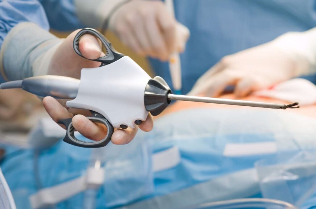
1. Laparoscopic graspers
Laparoscopic graspers are specialized instruments designed for minimally invasive surgery (laparoscopy). They allow surgeons to grip, hold, manipulate, and move tissues and organs through small incisions. Laparoscopic graspers consist of long, slender shafts with a handle on the proximal end that controls opposing jaws at the distal end.
Laparoscopic graspers serve as extensions of the surgeon’s hands during minimally invasive surgeries like cholecystectomies and appendectomies. These instruments can further improve surgeon performance by providing haptic feedback to surgeons – tactile sensations that help surgeons accurately assess tissue resistance, apply the right amount of force, and manipulate delicate structures with accuracy. This feature lets surgeons experience a more intuitive sense of touch, improving precision and reducing the risk of tissue damage.
2. Endoscopic staplers
Endoscopic staplers are surgical instruments that place rows of titanium staples on tissues while simultaneously cutting the tissues between the staple lines. These highly specialized instruments feature articulating heads that can rotate in multiple directions. Modern staplers use staple heights that adjust to different tissue thicknesses while maintaining the staple line.
Endoscopic staplers have proven especially useful for diverticulotomies, minimally invasive procedures for treating conditions like Zenker’s diverticulum. These specialized devices allow surgeons to precisely cut and simultaneously staple the diverticular pouch. A study from the World Journal of Gastroenterology found that laparoscopic graspers are safe instruments that lower the risk of bleeding and perforation during diverticulotomy.
3. Harmonic scalpels
Harmonic scalpels use ultrasonic waves to cut and cauterize (burn and close off) tissues. The scalpel is able to cut through tissue by converting electrical energy into mechanical vibration or waves. The generated waves have high frequencies in the range of 55.5 kHz that coagulate blood vessels with precision and denature tissue proteins.
Harmonic scalpels are commonly used during thyroid gland removal surgeries (thyroidectomies). One study found that using harmonic scalpels during thyroidectomy reduces operative time and is as safe as the conventional clamp-and-tie hemostasis techniques with regard to complications such as post-operative voice changes and bleeding.
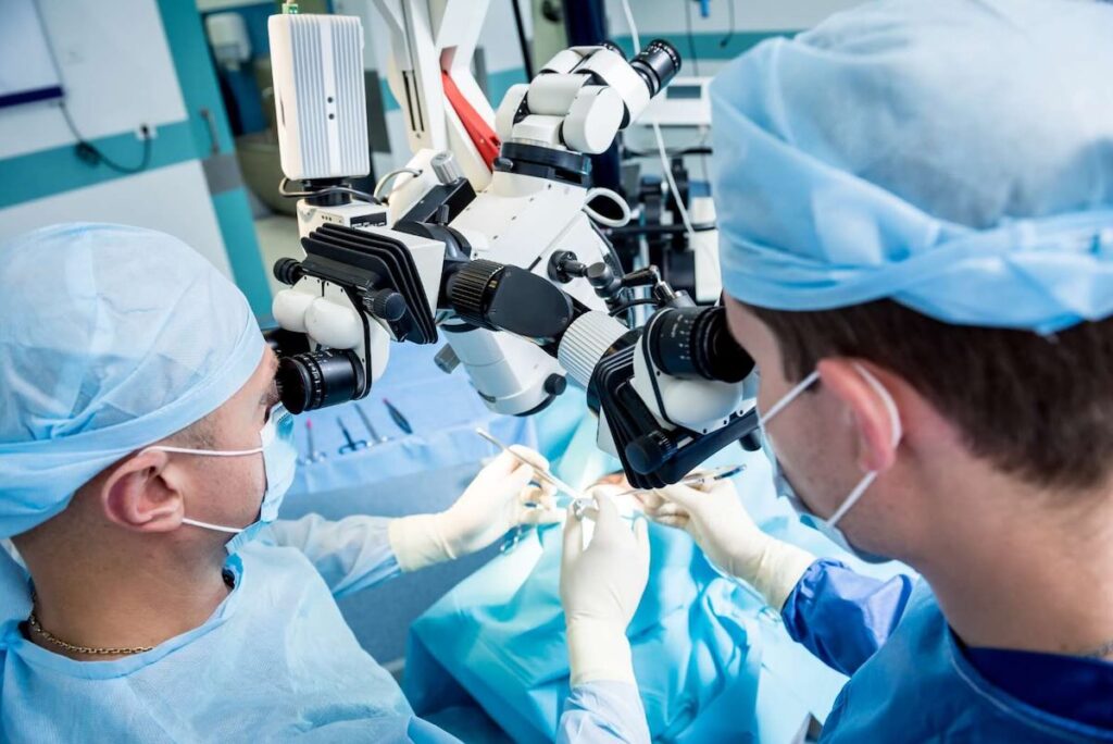
4. Surgical microscopes
Surgical or operating microscopes are optical instruments that provide magnification, visualization, and illumination of the surgical field. They are specialized for procedures requiring a high level of precision, such as:
- Brain tumor resection surgeries
- Nerve repairs and grafting
- Microvascular bypass procedures
- Carotid endarterectomies
- Microsurgical free tissue transfers
- Endodontic surgeries
Modern surgical microscopes feature LED illumination, integrated 3D and 4K video systems as well as apochromatic lenses, which minimize chromatic and spherical aberration.
Using a surgical microscope can reduce trauma and hospitalization time. Brain and spinal cord surgeries benefit the most from these specialized devices. The high magnification that the operating microscope helps surgeons remove tumors, manipulate tiny vessels, and access narrow spaces within the spinal canal with precision.
5. Handheld retractors
Handheld retractors are designed to hold back tissues and organs to improve surgical field visibility and access. They are typically small and, thus, operable by a single hand. Handheld retractors come in handy, especially for surgeries requiring precise incisions, such as tracheotomies and dental surgery.
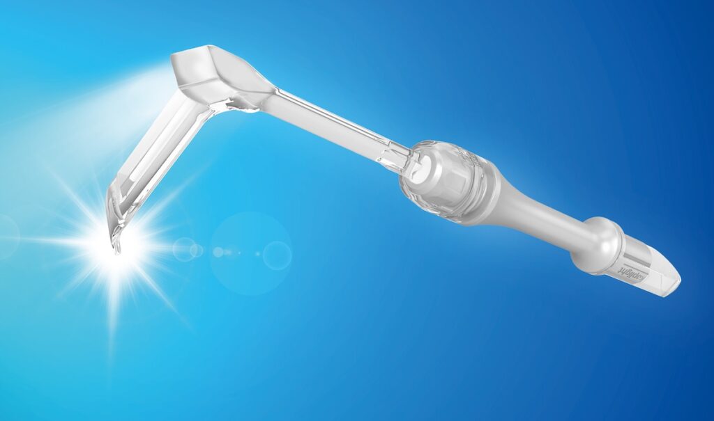
Modern retractors are available in various configurations including ribbon, rake, and malleable types. The Senn retractor, for instance, has three prongs that can allow for precise movements during precise surgeries, such as hand surgeries. In contrast, broader retractors like the Deaver retractor have curved blades that maintain steady retraction during long procedures.
Handheld retractors may be variants with integrated lighting. These devices are known as “lighted retractors”. They supplement the lighting from overhead systems by providing focused illumination of the surgical field.
6. Electrocautery devices
Electrocautery devices apply high-frequency electricity to biological tissue, generating heat that cuts tissue and coagulates blood vessels. Depending on the number of electrodes they use, these instruments can be monopolar (one electrode) or bipolar (two electrodes). Their power settings correspond to the tissue and surgery type.
Monopolar energy suits large surgical fields and procedures that dissect deep tissues, such as abdominal or orthopedic surgeries. In contrast, bipolar electrocautery is reserved for delicate surgeries where the stakes of collateral damage are high. Examples of such surgeries are neck surgeries or eye surgeries.
7. Bone saws
Bone saws or cutters are bone-cutting instruments. They have designs that can penetrate through hard, bony tissue with minimal heat generation. Bone saws are common instruments in highly invasive surgeries, such as orthopedic surgery, cardiothoracic surgery and neurosurgery. Examples of bone saw types include reciprocating cutters, unpowered saws, such as the Gigli saw, and costotomes.
In orthopedic joint replacement procedures, oscillating saws create precise femoral and tibial cuts that determine prosthesis alignment. In cranial procedures, craniotomes (specialized circular saws) create bone flaps for neurosurgical access. Costotomes, however, are specialized for cutting ribs. They are commonly used in thoracic and cardiothoracic surgeries.
A study by Minnaard et al. described novel bone cutters that can perform minimally invasive bone cuts. This method uses a steerable wire saw placement device that places a saw around the bone and cuts it without affecting surrounding tissues.
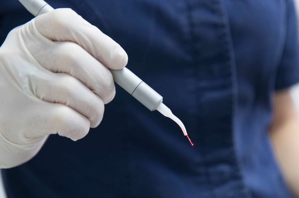
8. Surgical lasers
Surgical lasers are devices that emit light at specific wavelengths to cut, ablate, or coagulate tissues with precision. They are commonly used during tissue ablation, vessel sealing, and tumor removal surgeries. Major types include CO2 lasers, diode lasers, and Nd:YAG lasers; each of these types has distinct penetration depths and wavelengths that determine their usefulness in different procedures.
CO2 lasers
CO2 lasers produce infrared light at high wavelengths (10,600 nm). They are commonly used in aesthetic surgery to remove skin lesions and scars as well as reduce wrinkles and acne. CO2 lasers also assist in tonsillectomy (tonsil removal), vocal cord polyp removal, and leukoplakia treatment.
Diode lasers
Diode lasers are primarily absorbed by hemoglobin and melanin, making them effective for soft tissue procedures. Urologists use diode lasers for prostatectomy (prostate removal surgery), kidney stone surgery, and urinary stricture treatment.
Nd:YAG lasers
Nd:YAG lasers emit focused light at a wavelength of 1064 nm that penetrates into deep tissues. Vascular surgeons often utilize Nd:YAG lasers to treat varicose veins and hemangiomas. In oncology, Nd:YAG lasers are commonly used for tumor removal surgeries, particularly in cases where tumors obstruct the airway.
9. Arthroscopic shavers
Arthroscopic shavers are specialized instruments designed for minimally invasive joint surgeries (arthroscopies). Surgeons use these devices to “clean” (debride) a ligament in preparation for the reconstruction of the ligament. Arthroscopic shavers contain a handpiece connected to a console that powers a cutting blade rotating rapidly.
Orthopedic surgeons use these specialized devices during procedures of major joints, such as the knee, hip, shoulder, and elbow. Interestingly, arthroscopic shavers can also serve as processors of connective tissue progenitor (CTP) cells for later use. In a study published in the Journal of Clinical Medicine, researchers found that surgeons can use shavers to collect CTP cells from subacromial bursal tissue and use them to repair rotator cuff tears.
10. Vascular clamps
Vascular clamps are surgical instruments that temporarily occlude blood vessels during surgical procedures without causing vessel damage. They are common in cardiovascular surgeries. Modern clamps have atraumatic jaws with pressure-controlled closing mechanisms. They may also feature silicone covers with designs specific to the diameters of the targeted blood vessels.
The DeBakey clamp, for instance, assists surgeons in performing coronary artery bypass grafting (CABG), aortic aneurysm repairs, and vascular anastomosis procedures. On the other hand, the Bulldog clamp—a small, spring-loaded clamp—temporarily occludes small- to medium-sized vessels in plastic surgery and neurosurgical procedures.
11. Neurosurgical drills
Neurosurgical or cranial drills are high-precision instruments. They are specifically designed for controlled bone drilling and removal during brain and spine surgeries. Neurosurgical drills rotate at speeds of 20,000-80,000 rpm. They contain diamond or carbide burrs and incorporate irrigation systems that deliver a cooling solution to prevent excessive tissue heating.
In spinal procedures, these specialized instruments allow for precise laminectomy and foraminotomy to decompress the spinal cord or nerves. Surgeons operating on the skull base use diamond burrs to reduce bone thickness before final removal.
Some modern drills have integrated image-guided systems that define precise bone-cutting paths. These systems use two-dimensional images from computerized tomography (CT) or magnetic resonance imaging (MRI) scans to produce a three-dimensional image of bones. This application may potentially reduce operative time and the risk of infections and blood loss.
