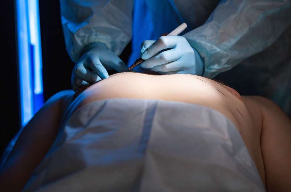
Various tools such as dyes, inks, sutures, and clips serve as skin and body tissue markers, enabling surgeons to precisely identify and delineate body tissues. And it’s crucial to mark body tissues during surgery to promote accuracy, safety, and effective communication.
Surgeons can correctly identify and mark operative sites, perform precise interventions, and navigate complex anatomical structures through tissue marking.
This article explores different ways of marking skin and tissue – their types, applications, and significance in enhancing surgical safety and efficacy.
Surgical dyes and stains
Surgical dyes and stains are used to color tissues and mark the borders of specimens removed during surgery.
Surgeons use various dyes and stains to create visual contrast, making it easier to identify and differentiate specific structures or areas of interest.
The most commonly used dyes and stains include Indocyanine green, Methylene blue, Patent blue, and Toluidine blue. These can be directly applied to the surface of body tissues for superficial marking or injected into tissues or vessels for deeper tissue staining and differentiation.
Using dyes and stains for tissue border marking extends to surgical pathology. Staining excised tissues with dyes helps surgical pathologists maintain the specimen’s correct orientation during an examination, so they can align it with its original positioning in the body. This aspect is especially significant in oncologic surgery, where accurate identification of “clear” or cancer cell-free margins is critical.
By employing dyes and stains to mark tissue borders, surgical pathologists can ensure precise evaluation of tissue samples. This helps determine whether the cancer is fully eradicated, or reoperation and removal of more tissue are needed.
Pros and cons
Using dyes and stains for tissue marking during surgery has advantages and disadvantages.
The main advantages of surgical dyes and inks include:
- Better visualization and localization. Dyes and stains provide visual contrast, improving the visibility and differentiation of specific structures or areas of interest within tissues. This enhances the surgeon’s ability to accurately identify and localize these structures, improving precision and minimizing the risk of errors during surgery.
- Facilitated orientation and communication. By using dyes and stains to mark whole margin planes rather than individual points on excised specimens, surgical pathologists can maintain the correct orientation during microscopic examination of residual tumor status. This approach reduces unnecessary re-operations, improves re-excision accuracy, and lowers cancer recurrence rates.
Fortunately, the potential disadvantages of tissue marking dyes and stains are relatively few. Some niche “cons” to consider include:
- Interference with histopathological evaluation. Certain dyes and inks used for tissue marking have been found to potentially interfere with immunohistochemical analysis and fluorescence in situ hybridization (FISH) techniques. This interference can impact the accurate interpretation of microscopic findings by pathologists, potentially affecting the diagnostic process.
- Variable fidelity. Some studies suggest that stains and dyes used for tissue marking may lose or change their intensity during special tissue processing. This variability can introduce errors in margin assessment and reporting, which may have serious implications for treatment decisions and patient outcomes.
Surgical ink and markers
Surgical ink is a specialized type of ink designed for use in surgical site marking pens or markers. It facilitates the precise marking of surgical sites during procedures such as incisions or percutaneous punctures.
It should be noted here that skin markers and body tissue markers are not the same thing. As the names imply, skin markers are for marking the body‘s surface, while body tissue markers are for marking within the body.
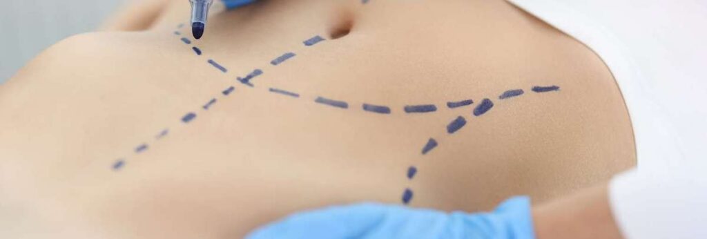
This marking helps indicate laterality (such as distinguishing between right and left), identifying specific levels (as in the case of spinal procedures), or marking multiple structures (like fingers or toes). Using surgical ink ensures accurate referencing and identification of intended surgical sites.
Surgical site marking pens use ink directly applied to the patient’s skin or tissue through a sterile disposable marker or pen. The desirable characteristics of surgical ink include:
- High visibility and resistance to smudging or fading during preoperative preparations
- Maintenance of clear visibility throughout the surgical procedure, providing a reliable reference point.
- Shouldn’t be so permanent as to last weeks or months after the surgical procedure.
The FDA classifies surgical site marking pens containing Gentian violet as Class 1 medical devices. And Gentian violet is the most common surgical marking ink. This water-based ink is known for its antibacterial, antifungal, and antihelminthic (antiparasitic) properties.
Yet, while such features suggest safety and effectiveness for direct skin use, there is opposition to Gentian violet.
Suggestions are that is carcinogenic, and this was a notable issue highlighted in California Proposition 65. This legislation attaches a warning to the use of this chemical. Gentian-free markers are beginning to emerge.
Despite the availability of these markers, in a pinch, non-toxic permanent ink markers or ballpoint pens, not specifically cleared by the FDA for direct skin use, have also been used for surgical site marking. This is improper use, but it’s known to occur.
Moreover, using a surgical skin marker to mark internal tissues can be both ineffective and unsafe. Dedicated body tissue markers are the best solution for marking inside the body cavity.
Surgical site marking is a vital element of the Universal Protocol for Preventing Wrong Site, Wrong Procedure, Wrong Person Surgery. These markings provide:
- A clear indication of the precise area of incision or operation
- Verification that the correct procedures are carried out on the correct sites
- Accurate placement and alignment of instruments during surgery
Pros and cons
The advantages of surgical site marking ink include increased accuracy and precision.
By applying pre-operative ink markings, the surgical team can readily identify the intended incision site or the specific area to be operated on. This proactive measure helps prevent errors such as wrong-site surgeries. Ultimately it also improves patient outcomes.
There are relatively few disadvantages of surgical site marking ink. The most relevant concerns include:
- The potential risk of cross-contamination and bacterial infection when pens are used on multiple patients.
- Risk for permanent “tattooing” and impaired wound healing if an incision is made directly over the ink markings.
- Deliberate or mistake use of skin markers and dyes for marking body tissues.
Sutures and ligatures
Suture and ligature markers are essentially the classic/original method for surgical tissue marking during procedures.
Sutures and ligatures can be used to mark important reference points, such as the borders of excised tissues, anatomic landmarks, sites of interest, or areas requiring further attention. In practice, however, these types of markers are only used to mark “poles” of excised tissues to orientate the specimens for pathologic examination.
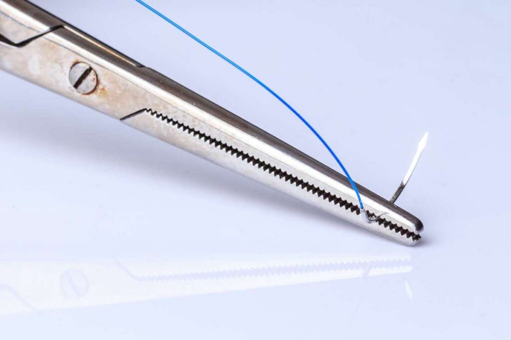
For example, in dermatologic surgery, potentially cancerous lesions are removed using an elliptical-shaped incision. Surgeons mark these ellipses with a suture at a defined pole while the specimen is still in situ. This helps them, and pathologists, with the correct orientation, monitoring, and further treatment.
Pros and cons
There are, in fact, few advantages to using sutures or ligatures as surgical markers.
- The most obvious benefit of sutures is their ready availability. Sutures and ligatures are commonly used in surgical practice and are always accessible.
- The second advantage of using these materials for demarcation is that they do not fade or lose visibility. As such, correctly placed suture or ligature markers will be easily visible to the surgical team.
- The last possible advantage is their ability to provide tactile feedback. This allows surgeons to visualize and feel for important landmarks or structures.
Marking tissues with sutures and ligatures also have some potential drawbacks to consider. The most pertinent concerns include:
- There are no universal guidelines to facilitate tissue marking with sutures or ligatures. This leads to variation in clinical practice. All of this translates to confusion and misinterpretation between surgeons and pathologists. One study showed discordance rates as high as 30–52% in identifying specimen margins between surgery and pathology when using suture markers.
- Inappropriate application, leading to tissue trauma, has also been reported when sutures are used to mark tissues. This can compromise the histological examination of specimens, leading to inappropriate re-interventions or missed diagnoses.
Metallic surgical clips
Metallic surgical clips serve as radiopaque implants used for tissue marking purposes. These devices are commonly employed by oncological surgeons and interventional radiologists before surgical procedures, particularly in breast biopsies.
The placement of these clips allows for various benefits:
- Localization of biopsied sites. Metallic clips act as tracking markers, aiding surgeons in locating previously biopsied areas. This is particularly valuable when pathology results indicate malignancy, as it enables accurate intra-operative tumor resection.
- Radiographic compatibility. Due to their radiographic visibility, metallic clips facilitate the demarcation of tumor borders and enable evaluation of tumor growth or shrinkage through imaging techniques like X-ray, CT scan, or MRI. This feature improves assessment of the tumor’s characteristics and response to treatment.
- Assurance of complete resection. Metallic clips assist pathologists in ensuring the removal of the lesion of interest. By identifying the region of interest in a mastectomy specimen, the clips help verify that the targeted tissue has been successfully excised.
Pros and cons
The advantages of metallic clips are mentioned above. There are also a few potential drawbacks to consider:
- A significant disadvantage of using a metal clip marker is that it remains permanently in tissues. As a result, many patients may decline to undergo a biopsy due to concerns about the clip remaining in the bodies indefinitely.
- Another drawback is the potential migration of metal clips away from the site of biopsy insertion shortly after they are deployed.
- The last disadvantage of these clips is their ability to alter the local magnetic field during MRI due to their ferromagnetic properties. This can potentially impact the sensitivity of MRI in detecting and monitoring cancer treatments.
Some surgeons come up with their own ways of marking
There are other reports of surgeons using techniques such as scraping or burning, such as with a scalpel, and using sterilized paper as a guide. Surgeons need reliable guidelines to isolate the right area and facilitate communication. Body tissue and skin markers are dedicated tools for the purpose, and the best ones are safe, sterile, and make a physician’s life easier.
