
Surgical lighting ensures proper illumination during surgery, improving surgical visibility and success. Methods of surgical lighting have advanced greatly in recent years. They now employ light-emitting diodes (LEDs) and include overhead lighting systems, wearable devices, and light-emitting instruments. This article explores the current practices and future prospects of surgical illumination.
Surgical lighting – What it is and why it’s necessary
Surgical lighting is specialized for illuminating and visualizing the patient and operating field during surgery.
Until the 19th century, surgeons operated using natural light, candles, and improvised light sources. Surgical lights became a reality with the discovery of electricity in 1879. The 20th-century revelation of halogen bulbs was a key turning point for surgical lighting.
Today, surgical lights use much more advanced technologies, such as LED. They come in various designs based on the surgeon’s needs and include:
- Surgical light systems
- Headlights and loupes
- Operating microscopes
- Lighted retractors
- In-cavity lighting
High-quality surgical illumination is necessary to improve surgery, protect the patient, and prevent complications, such as:
- Eye, neck, and back strain
- Distractions
- Surgical site infections
- Burns and fires
- Malpractice claims
- Increased surgery time and costs
Optimal surgical lighting is nimble, sterile, and easy-to-use. It offers clear illumination by balancing brightness and colors and managing shadows, glare, and heat. These characteristics must follow the International Electrotechnical Commission (ICE) 60601-2-41 standard, which specifies essential safety and performance requirements of surgical luminaires and luminaires for diagnosis.
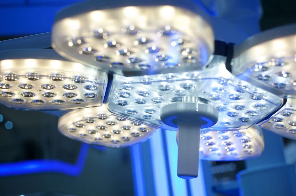
Surgical lighting systems / overhead lighting
A surgical lighting system (SLS) is the most commonly used surgical lighting method, as SLSs are proven to be stable and reliable for open surgery. They involve ceiling- or wall-mounted luminaires or lights in the operating room, providing overhead illumination during surgery.
The greatest advantage of an SLS is its ability to illuminate the surgical field from different angles. Its most notable disadvantage is that it requires manual repositioning. On average, light adjustments occur every 7.5 minutes during surgery, and 97% of the time, surgeons must pause the surgery to adjust the light themselves.
Sometimes, additional staff members must scrub in to help move the light when needed. An SLS may also be out of reach at times or cause accidental collisions, prolonging the surgery and distracting the surgeons.
The majority of overhead surgical lights contain halogen, LED, or xenon lamps.
Halogen bulbs
Halogen bulbs are common components of surgical lighting systems, headlights, and operating microscopes. They represent incandescent light bulbs filled with halogen gas that produce light when heated. Halogen bulbs produce a warm, yellowish color and last for 1,000–3,000 total hours (PDF link). Operating rooms must have heat protection filters and backup lights installed or available, as halogen bulbs might die abruptly, sometimes even during surgery.
Although halogen lights have been used in operating rooms for decades, they have deficiencies. They don’t provide efficient illumination, are costly in the long run, and cause contact burns. They may also damage the skin and eyes by emitting non-visible infrared and ultraviolet radiation.
LED bulbs
LED bulbs are largely replacing halogen bulbs for surgical lighting. They are more efficient, more durable, and safer than their halogen counterparts. Depending on their use, LED bulbs can last for up to 60,000 hours.
LED bulbs emit various colors, such as red, yellow, green, blue, or white, without causing heat risks. The color is typically determined by the type of material that makes up the bulb.
Colors can be warm (yellowish, pinkish) or cold (bluish) based on their temperature in Kelvin (K). Different colors affect eye strain, tissue color, and contrast differently. Colder colors (higher on the Kelvin scale) are similar to sunlight and allow for more precision during surgery, whereas warmer colors (lower on the Kelvin scale) are more appropriate for longer surgeries as they put less strain on the eyes.
Recent high-flux LED lights have remarkable luminous capacities of 100 lumens per watt (lm/W) and higher.
Xenon bulbs
Xenon bulbs produce a bright, cool, white light that mimics natural light. They offer better contrast and color balances than traditional halogen bulbs, which is why they are commonly used for wearable surgical devices during precise surgery, such as neurosurgery.
Xenon and LED lights can provide a tremendous brightness level of 220,000+ lux at an average distance of 16 inches. Most other surgical lights do not exceed 160,000 lux.
Xenon bulbs are not as energy-efficient or durable as LED bulbs. They tend to degrade early, lasting for up to 1,000 hours. Of all other light types, xenon light generates the most noise and heat. It is most often associated with intraoperative burns.
Headlights and other wearable lighting
Headlights or headlamps are wearable surgical lighting devices. They are typically made of a band that fits around the head and a frontal LED or xenon light source. The light source can be battery-powered or receive continuous power from a connected fiber-optic cable.
Headlights eliminate shadows and provide light that focuses on a specific target according to the surgeon’s attention and position. They are used for surgeries involving narrow, lateral, or deep surgical sites.
That said, headlights are associated with some unwanted effects. They can be heavy and uncomfortable to wear for prolonged periods, causing head and neck strain, which might progress to chronic musculoskeletal disorders. Their cables may also cause tripping accidents in the operating room.
Newer headlight designs aim to be cordless, lightweight, and ergonomic.
Illuminated surgical loupes
Surgical loupes are magnifying surgical lighting devices attached to the lenses or frames of surgical glasses. Surgeons use surgical loupes to visualize and magnify small internal structures. They are especially useful for precision surgeries, such as thyroid removal surgeries.

Loupes often contain a small lamp attached to the central part of the frames to illuminate the surgical site. Recent findings show that combining magnifying loupes with shadowless lamps and filtering eyeglasses protects the surgeon from light toxicity.
Operating microscopes
Operating or surgical microscopes are exclusive to microsurgery. They are unique lighting devices, allowing for a broad field of view with a three-dimensional magnification of structures ranging from 2.9x to 62x. They provide surgeons with clear illumination, binocular vision, and a personalized user experience.
The main downside of operating microscopes is the risk of burns associated with their use. Operating microscopes transfer large amounts of heat to tissues, potentially damaging them. Small blood vessels, the inner ear, and intraocular structures are especially vulnerable.
Current recommendations instruct surgeons to use the lowest possible setting that still provides a clear view of the operating field.
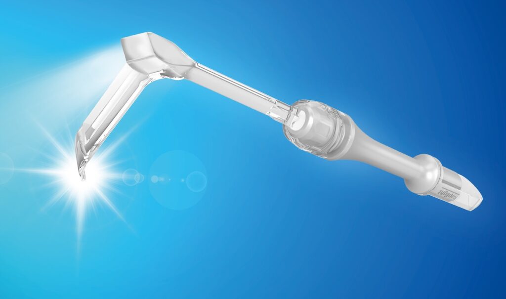
Lighted retractors
Lighted retractors are surgical instruments that:
- Keep the edges of a surgical wound separate
- Hold internal tissue away to provide better surgical access
- Illuminate surgical cavities with the help of an attached lamp
Lighted retractors can be handheld or self-retained. They may be cordless or operate using a fiber-optic cord. They provide light near the tissues, improving visibility and avoiding shadows or glares. Thus, in some ways, they may be superior to overhead lighting systems for illuminating body cavities.
Some lighted retractors can also transilluminate tissues. This means that they can determine the composition and density of tissues based on how the tissues diffuse light. Surgeons use tissue transillumination to differentiate between normal and abnormal tissues in cavities, such as during laparoscopic cholecystectomy (gall bladder removal minimally invasive surgery).
Handheld lighted retractors are easy to maneuver but occupy a surgeon’s or assistant’s free hand. They are smaller in size, not very durable, and unable to retract larger areas like the sternal region effectively. Conventional metal retractors often better meet such needs.
In-cavity surgical lighting
In-cavity surgical lighting provides illumination of deep and narrow surgical cavities. This is why it’s commonly used in minimally invasive surgery. An in-cavity lighting device is fixed outside of the sterile field, much like a surgical lighting system.
In-cavity surgical lighting can be coupled with in-cavity imaging for laparoscopic surgery – minimally invasive surgery of the abdominal cavity using small incisions and long, tubular instruments.
A problem with this particular technique is that it can generate direct heat, potentially harming tissue. To reduce direct tissue injury, in-cavity lighting can be used with fiber-optic cables.
Fiber-optic cables transmit light over a long distance without disrupting visibility. As mentioned, they assist in-cavity lighting, lighted retractors, and headlights. Fiber-optic cables consist of one of two materials:
- Polymers: affordable, cool, relatively safe
- Glass: provides better light transmission but is more expensive than polymers
While fiber-optic cables reduce the amount of direct heat generated, they can still heat up quickly themselves, giving rise to eventual contact burns, as mentioned. The cables may also become entangled, potentially causing tripping injuries.
Automated lighting systems
Lately, scientists have been exploring the potential of automated surgical illumination (PDF link), which would eliminate the need to manually adjust lights during surgery.
An automated, “touchless” lighting system uses artificial intelligence, tracking or gesture-based systems, or thermal imaging to illuminate the surgical field effectively at all times.
Automating surgical illumination may have the following benefits:
- Reduction of stoppages to manually adjust surgical lights
- Elimination of unnecessary contact with the surgical lights
- Prevention of collisions and mechanical problems
These effects lead to enhanced concentration levels among surgeons and operating room staff. They also avoid infections and other known complications. Thus, automated systems could significantly improve surgical performance and outcomes in the near future.
